When it comes to observing microscopic specimens, one of the most common techniques is preparing a wet mount. As an experienced microbiologist, I have perfected the art of creating clear and detailed wet mount slides. In this article, I’ll walk you through the step-by-step process of preparing a wet mount specimen for optimal viewing. Whether you’re a student conducting a biology experiment or a curious enthusiast exploring the wonders of the microscopic world, mastering this technique is essential for obtaining accurate and captivating observations. So, let’s dive in and uncover the secrets of creating a perfect wet mount slide!
When Preparing A Wet Mount Specimen For Viewing, The Specimen Should Be Covered With
To start preparing a wet mount specimen, you will need a microscope slide. This is a small rectangular piece of glass or plastic that serves as a platform for mounting and observing the specimen. Make sure the slide is clean and free from any dust or debris to ensure clear observations.
Cover Slip
When preparing a wet mount specimen for viewing, it is important to cover it with a cover slip. This is a thin, transparent piece of glass or plastic that is placed over the specimen on the microscope slide. The cover slip helps protect the specimen and prevents it from drying out. Additionally, it allows light to pass through the specimen, enabling clear and detailed observations.
Specimen
Of course, you’ll need a specimen to prepare a wet mount. This can be any microscopic material or organism that you want to observe. Whether it’s a plant cell, a drop of pond water, or a microorganism, ensure that the specimen is clean and properly prepared before placing it on the microscope slide.
Dropper or Pipette
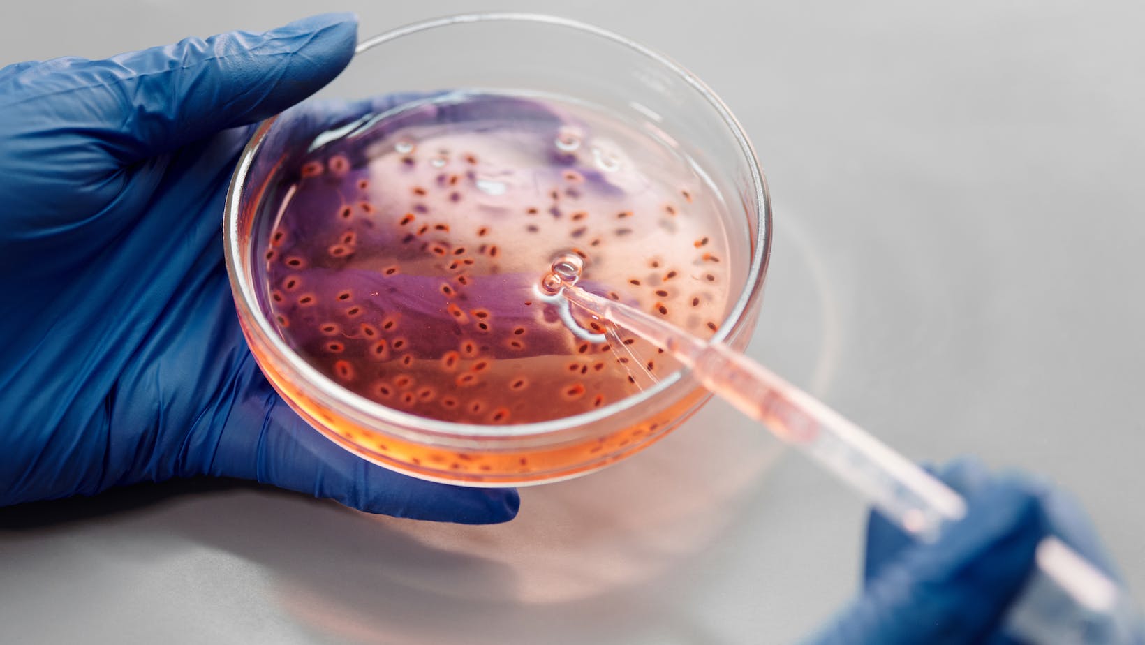
To add the necessary liquid to the slide and create the wet mount, you’ll need a dropper or pipette. This allows you to carefully add a small amount of water or mounting medium to the slide without disturbing the specimen. Precision is key here, as adding too much or too little liquid can affect the clarity of your observations.
Water or Mounting Medium
When preparing a wet mount specimen, you can use either water or a specialized mounting medium. Water is a common choice as it is readily available and can provide sufficient clarity for many specimens. However, for certain specimens or specific research purposes, a mounting medium may be necessary. The mounting medium helps preserve the specimen and provides better refractive index matching, resulting in improved image quality.
Remember, when preparing a wet mount specimen for viewing, make sure to cover it with a cover slip to protect it and ensure clear observations. Use a clean microscope slide, a dropper or pipette for precision, and choose between water or a mounting medium depending on your specific needs.
Steps for Preparing a Wet Mount Specimen
When preparing a wet mount specimen for viewing, the first step is to place a drop of water on the microscope slide. This water droplet will act as a medium for holding the specimen in place. It is important to use a clean slide to avoid any contamination that may interfere with the observation.
Transfer the specimen onto the water droplet
Next, carefully transfer the specimen onto the water droplet using a dropper or pipette. This ensures that the specimen is properly immersed in the water and is evenly spread out. When choosing the specimen, make sure it is covered with any necessary substances or materials that are required for observation.

Gently place the cover slip on top of the specimen
To protect the specimen and ensure clear observations, gently place a cover slip on top of the specimen. The cover slip should be carefully positioned at a slight angle, allowing it to gradually come into contact with the water. This technique helps to minimize the formation of air bubbles, which can interfere with the view.
Conclusion
Preparing a wet mount specimen for viewing under a microscope requires attention to detail and precision. By following the step-by-step instructions outlined in this article, you can ensure that your specimen is properly prepared and ready for observation.
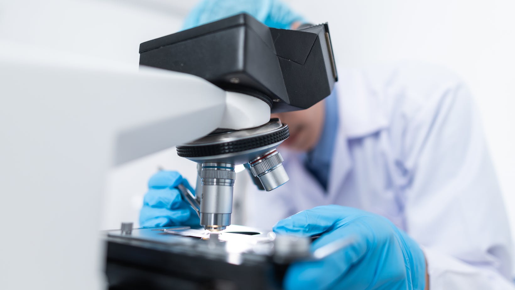
Once the wet mount specimen is prepared, it’s time to place it under the microscope and carefully observe any important details or observations. By taking note of these observations, you can further your understanding of the specimen and its characteristics.

